
¿Cómo se interviene quirúrgicamente el queratocono?
¿Qué es el queratocono?
La córnea es la pared transparente anterior del ojo que actúa como lente natural. El queratocono es una enfermedad que altera la forma normal de la córnea y provoca una deformación progresiva de ésta que en estadios finales llega a tomar una forma de cono, con lo que se altera progresivamente la calidad de la visión del ojo afectado.
¿Qué causa el queratocono?
Es una enfermedad con carácter hereditario y su penetrancia es variable, es decir, puede afectar minimamente o agresivamente variando su intensidad no solo entre paciente y paciente sino entre ambos ojos (se presenta de manera bilateral y asimétrica).
El proceso depende de la edad del paciente y el inicio de los síntomas. Habitualmente cuanto más joven es el paciente y más precoz es el inicio de queratocono, más rápida es la progresión.
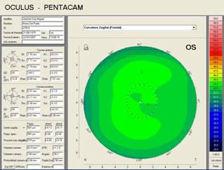
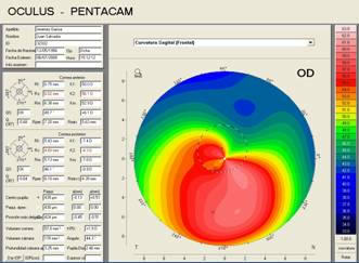
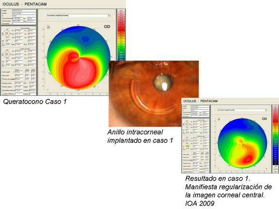
Aquí se observan dos imágenes de sendas topografías corneales para el diagnóstico del queratocono. Los tonos verdes de la imagen de la izquierda corresponden a una córnea normal En la imagen de la derecha se aprecian intensos tonos rojos que indican la presencia de un queratocono.
¿Cómo se interviene quirúrgicamente el queratocono?
Cuando la enfermedad esta poco avanzada el paciente puede usar gafas. Cuando la enfermedad progresa, la disminución de espesor y deformación de la córnea causa un astigmatismo irregular que no puede ya ser corregido adecuadamente con gafas y el paciente aqueja que nunca consigue ver del todo bien con ellas.
En estos casos se usan lentes de contacto rígidas que, si bien mejoran la visión, no detienen la progresión del queratocono. La intervención quirúrgica del queratocono es la única manera de detener la deformación progresiva de la córnea y, en muchos casos, de mejorar la visión. El diagnostico precoz influye decisivamente en el buen resultado del tratamiento.
¿Cómo se interviene quirúrgicamente el queratocono?
El Instituto Oftalmológico Amigó es el único centro de la provincia que utiliza el avanzado láser de femtosegundo para la realización de la cirugía. Esto significa que gran parte de la cirugía se realiza mediante laser guiado por ordenador, de esta manera se elimina en gran parte la parte manual y “artesanal” de la cirugía, obteniendo resultados predecibles, reproducibles, con menos complicaciones y, en definitiva, una técnica mucho más segura.
Existen diferentes tratamientos, y la elección del más adecuado dependerá de lo avanzada que esté la enfermedad y del grado de afectación visual.
- Cross-Linking corneal: Cuando se detecta que la enfermedad esta avanzando (progresando), el método de endurecimiento corneal denominado cross-linking es hoy por hoy la técnica mas indicada y con eficacia demostrada para detener el empeoramiento progresivo de la visión.
- Anillos intracorneales: esta técnica se emplea cuando la calidad visual del paciente es mala y no puede mejorarse eficazmente con gafas o lentes de contacto. Puede asociarse o no al Cross-linking. Por ser una técnica relativamente sencilla y de escasos riesgos, constituye un gran avance en el moderno tratamiento del queratocono que junto con el cross-linking ha disminuido muy significativamente la necesidad de un trasplante de cornea que hasta ahora era la única y dramática posibilidad de mejorar la visión en los casos avanzados. La incorporación del láser de femtosegundo al Instituto Oftalmológico Amigó ha supuesto una revolución igualmente en los implantes de anillos intraestromales para queratocono, simplificando los procesos y optimizando los resultados. Al igual que en la cirugía láser, el uso del láser de femtosegundo significa una disminución en los riesgos de la cirugía, dado que ya no es necesario utilizar bisturí o realizar el túnel corneal manualmente sino que es creado con una precisión inigualable mediante el láser.
- Trasplante de córnea: Cuando se ha dejado que la enfermedad avance en exceso, ni el cross-linking ni los anillos intracorneales son ya efectivos y la única solución es el injerto de una nueva córnea, proceso realmente más complejo y arriesgado.
A continuación le respondemos a alguna de las preguntas más frecuentes sobre los dos tratamientos que realizamos en el Instituto Oftalmológico Amigó, el cross-linking y los anillos intracorneales:
EL CROSS LINKING
Consiste en la aplicación de una sustancia endurecedora (Riboflavina) sobre la cornea enferma al tiempo que se ilumina con luz ultravioleta para obtener un aumento del entrecruzamiento (Cross-Linking) de las fibrillas de colágeno que conforman el tejido corneal, deteniendo así la deformación corneal y pudiendo llegar a mejorar la calidad visual.
Es una técnica indolora que solo precisa de anestesia en forma de gotas, tras lo cual el paciente acostado debe de mirar hacia una fuente de luz al tiempo que goteamos la Riboflavina sobre el ojo enfermo. El tratamiento no es corto pues llega a durar en total algo más de una hora.
LOS ANILLOS INTRACORNEALES
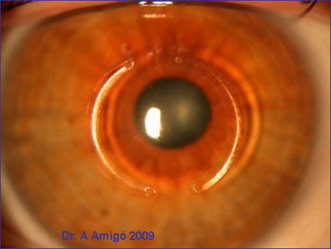
Es un tratamiento quirúrgico que se realiza sin penetrar al interior del ojo y respetando la cornea del paciente a diferencia del trasplante corneal. Se implantan uno o dos segmentos semicirculares rígidos en el espesor de la córnea que actúan tensando y regularizando el centro de la misma mejorando así la calidad de la visión deteriorada por el queratocono.
Los anillos están hechos de Perspex, se trata de un material acrílico que ha sido utilizado durante mas de 25 años en la fabricación de lentes intraoculares y que ha demostrado ser perfectamente tolerado por el organismo, sin riesgo de rechazo. Este tratamiento quirúrgico con anillos intracorneales es con mucho más seguro y menos traumático que el trasplante de córnea para casos de queratocono no excesivamente avanzado.
El grado de mejoría depende de lo avanzado del queratocono pues los pacientes operados en estadios iniciales son los más beneficiados con la cirugía con un porcentaje de éxito significativamente muy alto (85-90%).La recuperación visual es rápida y suele notarse ya desde los primeros días pero ha de recordarse que posiblemente sea necesario el uso de gafas o lentes de contacto para completar la mejoría visual.
Hoy en día la cirugía del queratocono con Cross Linking y/o anillos intracorneales es nuestra técnica de elección para el tratamiento quirúrgico del queratocono dada la rápida recuperación y las escasas complicaciones.
Conoce más aspectos sobre el queratocono en nuestro blog.