
Diagnóstico del glaucoma
¿Cómo se detecta si tengo principio o no de glaucoma?
Con el paso de los años observamos que la forma con mucho más frecuente de glaucoma (glaucoma crónico de ángulo abierto) generalmente no es advertida por el paciente hasta que se llega a perder visión central (la visión que por ejemplo usamos cuando miramos a los ojos de una persona).
Hemos visto repetidamente que la enfermedad pasa desapercibida porque el ojo menos afectado suele “trabajar” por el otro. También porque la gente suele prestar mas atención a la parte central de la visión, que no está afectada hasta que la enfermedad esta ya muy avanzada.
Finalmente, mucha gente asume que la pérdida de visión periférica (por los lados) es normal con la edad, cosa que en absoluto es así. Por ello actualmente aconsejamos a todo paciente de más de 40 años que debe ser controlado por el oftalmólogo al menos cada dos años. Como veremos a continuación, algunos exámenes son especialmente importantes para el diagnóstico del glaucoma.
¿Cómo detectamos y controlamos el glaucoma en el IOA?
En el Instituto oftalmológico Amigó examinamos tres condiciones principales para llegar al diagnóstico de glaucoma:
1. Determinar la presión intraocular
|
En primer lugar determinamos si la presión intraocular está elevada lo que se asociaría con un mayor riesgo de desarrollar la enfermedad.
Además, sabemos que las posibilidades de conservar la visión están relacionadas con una disminución significativa de la presión intraocular. La medición de la presión ocular o tonometría se realiza habitualmente con el llamado “examen de la luz azul” y que se trata de un aparato que se apoya en el ojo (tonómetro de aplanación de Goldman). Si bien en general este método indoloro es relativamente exacto, actualmente se sabe que en algunos pacientes puede ser erróneo al no tenerse en cuenta el grosor de la córnea (la lente transparente en la superficie anterior del ojo) grosor que influye directamente en la exactitud de los métodos de medida de la presión intraocular. Por esta razón desde 2008 nuestro Instituto dispone y utiliza de forma sistemática el ORA (OCULAR REICHELT ANALYZER), en todo paciente de mas de 40 años que nos consulta. Se trata de un equipo sofisticado que además de permitir obtener el valor real de la presión intraocular teniendo en cuenta el grosor de cada córnea, nos indica también la calidad del tejido ocular y con ello la predisposición a padecer glaucoma de cada paciente. ORA es diferente de la tonometría de no contacto con chorro de aire, que es otro método, utilizado frecuentemente en las ópticas, para medir la presión ocular. Utiliza un instrumento que “sopla” sobre el ojo y toma la medición de forma indolora. Considerada menos exacta que la tonometría de aplanación es utilizada con frecuencia para el screening del glaucoma. |
 Medicion “clásica” de la presión ocular con tonómetro de aplanación
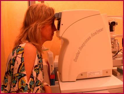 El ORA del IOA es probablemente el método actual más exacto para la medición real de la presion ocular.
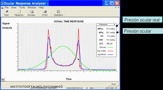 Diagrama resultado de una medición de la presion ocular con el ORA en un caso de glaucoma. Obsérvese como la presión ocular real de 31.4 es más alta que la presión ocular 29.1 que daría al obtenerse con métodos clásicos. |
2. Determinar el daño en el nervio óptico
El siguiente examen que utilizamos en nuestro Instituto es para determinar si existe ya daño en el nervio óptico o no, dado que el glaucoma se caracteriza por ir dañando progresivamente el tejido de este nervio que sirve para transmitir la visión del ojo hasta el cerebro.
El método actual mas sensible para detectar daños iniciales es el que realizamos mediante un Escáner del nervio óptico (OCT) y sus fibras, lo que nos permite saber si tiene usted un nervio óptico normal o no para su edad, si existe ya daño en el nervio y en que cantidad o si este daño progresa con el tiempo o no.
|
En la primera imagen de la derecha (») se observa como se obtiene un escáner del nervio óptico que nos permite detectar y tratar el glaucoma desde sus inicios, cuando con otros métodos aun no es posible detectarlo.
Se trata de un método sencillo e indoloro que utiliza un sofisticado haz de luz que permite en unos minutos y en la misma consulta estudiar el grado de salud del nervio óptico. La gran ventaja de este nuevo método del escáner de nervio óptico es que se anticipa a otros métodos al uso, como la clásica campimetría visual, lo que nos permite controlar el glaucoma antes de que el daño en la visión este avanzado o incluso antes de que llegue a producirse. En la segunda imagen de la derecha (») se observa el circulo en color verde junto a N.O. derecho (flecha superior), color que nos indica la total ausencia de daño. Mientras que el circulo con sectores ya en rojo junto a N.O. izquierdo (flecha inferior), nos señala que el glaucoma ya ha dañado seriamente a varias partes del nervio óptico en solo un ojo de este paciente. |
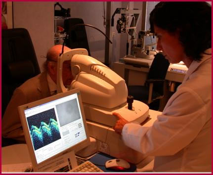 Realización de una OCT (Tomografía de Coherencia Óptica) en el IOA. |
 Resultado de una medición mediante OCT. |
3. Medición del campo visual
|
Una vez detectado el daño en el nervio óptico mediante la OCT, el siguiente método que utilizamos es la medición del campo de la visión de cada ojo campimetría o perimetría visual computarizada)
El campo visual o campimetría, nos confirma la presencia de daño en el nervio óptico, aunque desgraciadamente solo cuando ya que existe un daño relativamente avanzado, por ello la OCT no es de gran valor en estadios iniciales. Sin embargo, una vez aparece el daño en el campo visual, este método es extremadamente útil para, mediante sofisticados programas estadísticos computarizados, establecer si la enfermedad esta detenida con el tratamiento o continúa progresando y el tratamiento debe ser modificado. Por esta razón es crucial que realicemos campimetrías periódicamente en todo paciente con glaucoma o sospecha de padecerlo. La disminución del campo visual que se aprecia en la fotografía de la derecha, es uno de los tres signos principales del glaucoma avanzado a resultas del daño progresivo del nervio óptico. Una vez dañado la pérdida visual no puede ya recuperarse. La disminución del campo visual que se aprecia en la fotografía de la derecha, es uno de los tres signos principales del glaucoma avanzado a resultas del daño progresivo del nervio óptico. Una vez dañado la pérdida visual no puede ya recuperarse. |
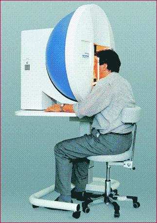 Campimetría visual computarizada en el estudio del glaucoma
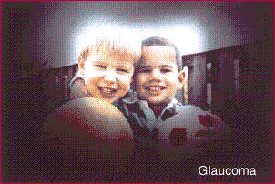 Visión con glaucoma |
Volver a
|
Volver a
|
Ir a tratamiento
|